The interest of SPF10 PCR-based methods for HPV DNA detection in FFPE samples
HPV and associated cancers: a global burden
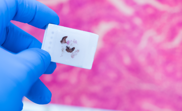
The past decades, prevalence studies have been done using FFPE (Formalin-fixed-paraffin-embedded) tissue specimens to investigate HPV association with a variety of clinical conditions e.g. cervical, anogenital and head and neck cancers, or oral cavity, laryngeal carcinoma, or anal, penile, and vulvar squamous cell carcinoma. The link between cervical cancer and HPV is well-established. Since 2017, direct HPV testing is recommended in the revised WHO/IARC classification to assess a correct diagnosis of the Head and Neck Squamous Cell Carcinoma (HNSCC).1
FFPE tissue: an important sample resource
FFPE tissues represent the most frequent form of tissue storage in pathology departments. These archival tissues are a potentially useful resource for epidemiological studies. Several studies have used FFPE specimens to investigate the correlation between HPV genotypes and histological classification, and/or to determine the prevalence of HPV in primary cancers.2-7
Fragmentation of DNA in FFPE samples
HPV detection and genotyping in FFPE samples is technically challenging due to poor DNA quality. Formalin fixation can cause DNA damage, including cross-linking and fragmentation.8
SPF10 PCR-based methods: most effective and most suitable
HPV DNA PCR methods amplifying shorter fragments of viral genome showed to be more sensitive and amenable for FFPE tissue. SPF10 PCR-based methods are mentioned as the most effective and most suitable for HPV DNA detection in FFPE samples because using a short HPV target region limits the risk of false-negative or invalid results.3,8-11,19
The article continues below.
INNO-LiPA HPV Genotyping Extra II on Formalin-Fixed-Paraffin-Embedded tissue
High sensitivity and specificity:
- Proven performance on cervical samples12
- The SPF10 PCR enables identification of HPV infections with low viral load and multiple HPV genotypes in one sample and one testrun12,13,14
- The small size SPF10 amplicon allows reliable amplification and accurate detection of HPV DNA also from FFPE samples5,6,14,20,21 and first-void urine15,16,17,18
- Compared to other HPV genotyping methods, INNO-LiPA is more likely to identify HPV genotypes in samples indicating low viral load and FFPE material containing multiple HPV genotypes11
Integrated controls for sample quality:
- Build-in human DNA control
- HPV control lines to confirm and detect the presence of a broad range of mucosal HPV genotypes
Prevention of co-amplification or amplicon carry-over:
- Build-in N-uracil glycosylase-based prevention minimizing the risk of PCR contamination and significantly reducing the possibility of false-positive HPV DNA results3
INNO-LiPA HPV GENOTYPING EXTRA II can play an important role in:
- Investigation of the prevalence of HPV and genotype distribution in different types of cancer 2,4-6,19-21
- Evaluation of vaccination trials and monitoring the impact of HPV vaccination (pre-and post-vaccination monitoring)15,18,19
- Epidemiological studies to investigate the prevalence and distribution of HPV types13,14
References:
- Westra W. et al. Head and neck pathology, 2017; 11:41-4
- Pretet JL. et al. Int J Cancer, 2008; 122:424-427
- Kocjan B. et al. Journal of Clinical Virology, 2016; 76:88-97S88-S97
- Sinno A. et al. Obstet Gynecol, 2014; 123:817-821
- Acuña G. et al. Modern Pathology, 2019; 32:621-626
- Fuglsang K. et al. Papillomavirus Res, 2019; 7:15-20
- Valmary-Degano S. et al. Human Pathology, 2013; 44:992-1002
- Steinau M. et al. J Mol Diagn, 2011; 13:377-381
- Tan SE. et al. J Clin Microbiol, 2010 ; 48 :1458-1460
- Querec T. et al. IPVC 2020 Book of Abstracts p.1013-1014
- Bicskei B. et al. 2020. J Cancer Sci Clin Ther, 2020; 4:349-364
- Xu L. et al. Int J Mol Sci, 2018; 19:2704
- Sohrabi A. et al. J Infect Public Health, 2017; 10:730-733
- Ahmadi S. et al. Asian Pac J Cancer Prev, 2017;18:3373-3377
- O’Leary MC. et al. Br J Cancer, 2011; 104:1221-1226
- Ducancelle A. et al. Arch Gynecol Obstet, 2014; 290:299-308
- Jannes G. et al. EUROGIN 2018 Poster P10-5
- Burroni E. et al. J Med Virol, 2015; 87:508-515
- Hillman R. et al. Int J Cancer, 2014; 135 :996-1001
- Dalla Libera LS. et al. J Oncol, 2019; Article ID 6018269
- Swangvaree SS. et al. Asian Pac J Cancer Prev, 2013; 14:1023-1026
Related articles

Advancing anal cancer prevention: Screening expansion and the emerging role of host-cell DNA methylation
Anal cancer incidence remains low in the general population but is significantly elevated in defined at-risk groups, including people living with HIV...
HPV genotyping in FFPE tissue: meeting the challenges of archival sample analysis
Human papillomavirus (HPV) is a necessary cause of cervical cancer and is increasingly recognized as an important etiological factor in a range of...

Clinical validation of the PreCursor-M AnoGYN test: Tailoring treatment of anal and vulvar cancer precursors
A major challenge for the correct implementation of the anal cancer screening guidelines is the generally limited availability of High-Resolution...

New trends in cervical cancer screening: Self-sampling would allow full molecular screening – great opportunity to increase attendance to cervical cancer screening
Cervical cancer screening is evolving, and one of the most promising innovations is full molecular self-screening. This method combines HPV testing...

EUROGIN 2025 – key takeaway messages
The EUROGIN 2025 conference, held at the Alfândega Congress Center in Porto, Portugal, focused on advancing scientific efforts to control HPV-related...

Updated anal cancer screening guidelines now provide a basis for expansion of screening to all at-risk populations
Screening guidelines for anal cancer prevention have traditionally focused on people with HIV, and focus on the detection of high-grade squamous...

EUROGIN 2024 – key takeaway messages
The annual EUROGIN conference covers a dense scientific program on the topic of Human papillomavirus (HPV) and its associated cancers, always with a...

Scientific poster – Inter- and intra-laboratory reproducibility of the AmpFire® HPV Screening 16/18/HR assay
This poster summarizes the investigation of the reproducibility (within a laboratory and between laboratories) of the AmpFire HPV Screening 16/18/HR...

A risk-based approach for post-treatment detection of recurrent CIN
This article describes how HPV genotyping and DNA methylation analysis can help discriminate women in need of re-treatment from those who can be...

Video: DNA methylation and improved detection of cervical pre-cancers in screening, diagnosis and post-treatment management
In this short video you will find out how DNA methylation plays a crucial role in cancer development. And how the PreCursor-M+ assay, measuring the...

Better care for pregnant women diagnosed with precancerous cervical lesions
Cervical cancer is the most common malignancy of the female reproductive tract during pregnancy occurring in 1 to 10 per 10 000 pregnancies with an...

Cervical cancer screening being reviewed – the role of distinguishing individual HPV genotypes
Cervical screening guidelines are being reviewed and changed in response to new evidence as it becomes available. At EU level the cancer screening...

Improved triage with FAM19A4/miR124-2 methylation analysis - clinical assurance thanks to objective and reliable results
Studies demonstrate FAM19A4/miR124-2 methylation can be considered as an alternative to cytology as follow-up test for hrHPV positive women given its...

EUROGIN 2023 - Shared challenges to the implementation of HPV screening and diagnostics from research to practice
The keynote lectures by prominent experts in their respective fields at the recent Eurogin 2023 Conference in Bilbao, Spain, once again gave a good...

Booklet: The Art of Accuracy - Improved triage with FAM19A4/miR124-2 methylation analysis
How does FAM19A4/miR124-2 methylation analysis improve hrHPV positive patient triage and how does it allow a wait-and-see policy? We have prepared an...

Webinar: First void urine & HPV genotyping - Two essential tools to monitor the impact of HPV vaccination programs
The enormous potential of a first void – or initial stream – urine sample becomes evident when we understand it’s composition. In fact, in the case of...

Interview: Urine self sampling could change the world of testing, not only for HPV but also for other oncology markers
Our own Rebecca Millecamps, Marketing Manager at Fujirebio, was interviewed for the podcast series 'Molecules, microbes and multiomics' about the...

INNO-LiPA® HPV Genotyping Extra II for use on self-collected first-void urine samples
HPV (Human Papilloma Virus) is a sexually transmitted virus. Certain genotypes of this virus are known to cause cancers in both men and women, but...


 Powered by Bioz